Are There Different Types Of Accessory Pathways
Lown, B. The syndrome of short P-R interval, normal QRS complex and paroxysmal rapid heart action. Circulation. vol. 5. 1952 May. pp. 693-706.
James, TN. Morphology of the human atrioventricular node, with remarks pertinent to its electrophysiology. Am Heart J. vol. 62. 1961. pp. 756-71.
Lev, M, Leffler, WB, Langendorf, R. Anatomic findings in a case of ventricular preexcitation terminating in complete atrioventricular block. Circulation. vol. 34. 1966. pp. 718-33.
Murdock, CJ, Leitch, JW, Teo, WS. Characteristics of accessory pathways exhibiting decremental conduction. Am J Cardiol. vol. 67. 1991. pp. 506-10.
Ross, DL, Uther, JB. Diagnosis of concealed accessory pathways in supraventricular tachycardia. Pacing Clin Electrophysiol. vol. 7. 1984. pp. 1069-85.
Anderson, RH, Becker, AE, Brechenmacher, C. Ventricular pre-excitation: a proposed nomenclature for its substrates. Eur J Cardiol. vol. 3. 1975. pp. 27-36.
Mahaim, I, Benatt, A. Nouvelles recherches sur les connections superieures de la branche du faisceau de His-Tawara avec cloison interventriculaire. Cardiologia. vol. 1. 1937. pp. 61
Also Check: Similar To Parkinsons
Taquicardias Generadas Fuera Del Circuito:
Son taquiarritmias generadas en las aurÃculas sin relación con el Wolff-Parkinson-White , que se trasmiten a los ventrÃculos a través de la vÃa accesoria. Al no tener la vÃa las propiedades de conducción lenta del nodo AV, puede provocar frecuencias ventriculares peligrosamente elevadas y generar fibrilación ventricular.
Atrial Fibrillation And Wpw
Patients with Wolff-Parkinson-White syndrome have an accessory pathway or a bypass tract that connects the electrical system of the atria directly to the ventricles, thereby allowing conduction to avoid passing through the AV node.
In normal individuals, when the sinus node creates an action potential it must pass through the AV node to get to the ventricles. When an accessory pathway is present, the sinus node action potential can pass through the bypass tract before the AV node, which causes the ventricles to become depolarized quickly. This is termed pre-excitation and results in a shortened PR interval on the ECG.
The combination of WPW and atrial fibrillation can potentially be fatal, especially if AV blocking agents are given . The medical treatment is procainamide, although electrical cardioversion is reasonable, especially if hemodynamically unstable.
In patients with WPW and atrial fibrillation, the erratic atrial action potentials can conduct through the accessory pathway very quickly . Therefore, WPW patients who develop atrial fibrillation have higher ventricular rates than those without WPW. If an AV blocking agent is given, fewer atrial action potentials will pass through the AV node and more will pass through the accessory pathway, paradoxically increasing the ventricular rate potentially causing ventricular fibrillation which is a fatal, hemodynamically unstable rhythm. Procainamide or electrical cardioversion is recommended in these situations.
You May Like: Last Stages Of Parkinson’s Before Death
Cleveland Clinic Heart Vascular & Thoracic Institute Cardiologists And Surgeons
Choosing a doctor to treat your abnormal heart rhythm depends on where you are in your diagnosis and treatment. The following Heart, Vascular & Thoracic Institute Sections and Departments treat patients with Arrhythmias:
- Section of Electrophysiology and Pacing: cardiology evaluation for medical management or electrophysiology procedures or devices – Call Cardiology Appointments at toll-free 800.223.2273, extension 4-6697 or request an appointment online.
- Department of Thoracic and Cardiovascular Surgery: surgery evaluation for surgical treatment for atrial fibrillation, epicardial lead placement, and in some cases if necessary, lead and device implantation and removal. For more information, please contact us.
- You may also use our MyConsult second opinion consultation using the Internet.
The Heart, Vascular & Thoracic Institute has specialized centers to treat certain populations of patients:
Which Congenital Heart Disease Is Associated With Pre

Left ventricular pre-excitation was recorded in 18 cases: 8 in the lateral zone, 5 in the anterior paraseptal and 5 in the posterior paraseptal zones. WPW and congenital heart disease: Out of 20 cases of Ebsteins anomaly, 5 cases of WPW were observed: 4 right posterior and 1 right lateral pre-excitations.
You May Like: Pfnca Wellness Programs
You May Like: Que Es La Enfermedad De Parkinson Y Sus Sintomas
How Is Wpw Diagnosed
WPW can only be diagnosed by reviewing an ECG . A holter or ambulatory monitor and exercise testing are also helpful in evaluating patients known to have WPW.
In the past, patients with WPW but without symptoms had been observed by a cardiologist for many years. Recently, new guidelines have been published for this group of patients. Your cardiologist may order a holter monitor or stress test to look for a persistent patter of WPW. If the WPW pattern persists, invasive electrophysiology testing is now recommended.
Your doctor will also ask you several questions:
- Do you have symptoms?
- Do you have a history of atrial fibrillation?
- Do you have a history of fainting?
- Do you have a history of sudden cardiac death or does anyone in your family?
- Are you a competitive athlete?
The results of your diagnostic tests and the answers to these questions will help guide your therapy.
How Is Wpw Different From Typical Avrt
The difference between this typical AVRT and the AVRT seen with WPW is that, in WPW, the accessory pathway is capable of conducting electrical impulses in both directions from the atrium to the ventricle as well as from the ventricle to the atrium.
As a result, during reentrant tachycardia in WPW, the electrical impulse is able to travel down the accessory pathway into the ventricles, then return to the atria through the AV node, then back down the accessory pathway to the ventricles again and it can keep repeating the same circuit. This is the opposite direction of travel than in patients with typical AVRT.
Read Also: Singing And Parkinson’s Disease
Why Wpw Is A Particular Problem
The ability of the accessory pathway in WPW to conduct electrical impulses from the atria into the ventricles is important for three reasons.
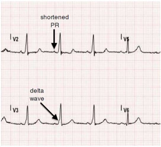
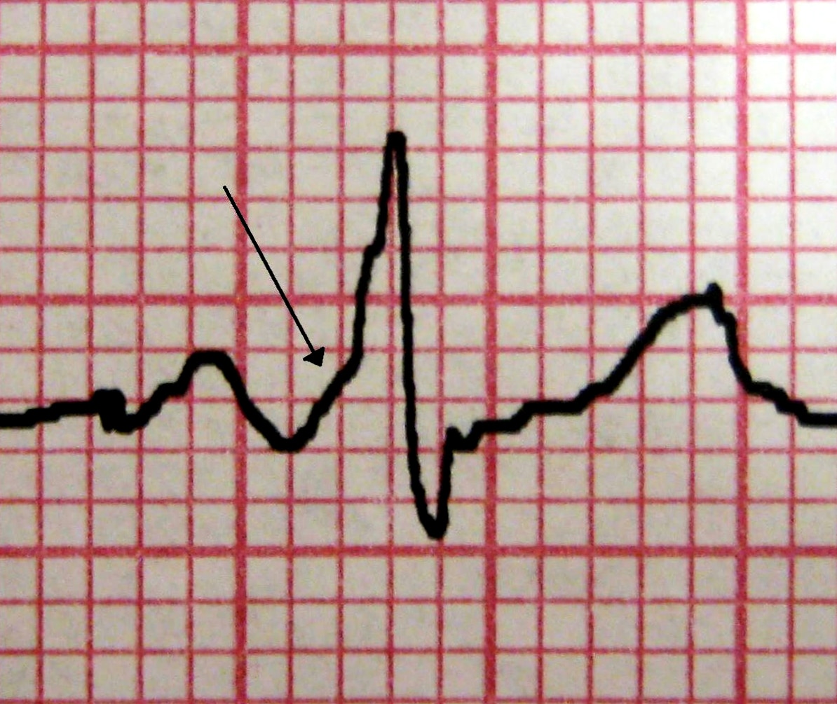
First, during normal sinus rhythm, the electrical impulse spreading across the atria reaches the ventricles both through the AV node and through the accessory pathway. This “dual” stimulation of the ventricles creates a distinguishing pattern on the ECG specifically, a “slurring” of the QRS complex which is referred to as a “delta wave.” Recognizing the presence of a delta wave on the ECG can help a doctor can make the diagnosis of WPW.
Second, during the AVRT seen with WPW, the electrical impulse is stimulating the ventricles solely through the accessory pathway . As a result, the QRS complex during tachycardia takes on an extremely abnormal shape, which is suggestive of ventricular tachycardia instead of SVT. Mistaking the AVRT caused by WPW for VT can create great confusion and unnecessary alarm on the part of medical personnel, and may lead to inappropriate therapy.
Third, if a patient with WPW should develop atrial fibrillation an arrhythmia in which the atria are generating electrical impulses at an extremely rapid rate those impulses can also travel down the accessory pathway and stimulate the ventricles at an also extremely rapid rate, leading to a dangerously fast heartbeat. So in patients with WPW, atrial fibrillation can become a life-threatening problem.
Localization Of Accessory Pathways
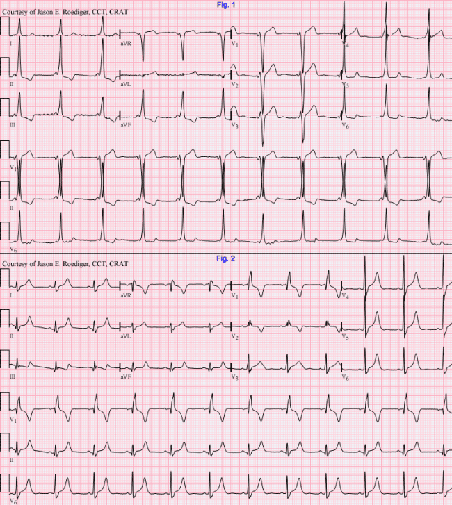
The location of the AP can often be determined through analysis of the spatial direction of the delta wave in the 12-lead ECG by reviewing the maximally preexcited QRS complexes. A general rule is that Q waves point away from the earliest site of ventricular activation, which is typically the insertion point of the bypass tract. The most common locations for APs, in decreasing order of frequency, are the left free wall, the posteroseptal and right free wall, and finally the midseptal and anteroseptal regions of the heart.
Several algorithms are available to predict the location of the AP. These algorithms may not be totally accurate because maximal preexcitation is needed, and usually the QRS in WPW pattern is a fusion between AV node and AP depolarization , precordial lead placement may vary, as well as chest shape and size and heart shape, size, and location.
A practical concept is that a negative delta wave usually signals the location of the AP, as follows:
- A negative delta wave in a left-side lead such as I and aVL indicates a left-side AP
- A negative delta in a right-side lead such as V1 predicts a right-side AP
- An isoelectric delta in V1 predicts an anteroseptal AP
- A negative delta in the inferior leads indicates a posteroseptal AP
- A positive delta in the inferior leads predicts an anteroseptal AP
A more specific algorithm for location of the AP, based on the polarity of the delta wave or first 40 ms of the QRS, predicts the following AP locations:
Don’t Miss: Adaptive Silverware For Parkinson’s
Electrocardiographic Signs Of Pre
As the stimulus originates from the sinus node, the P wave will be normal.
In pre-excitation, ventricles depolarize from two different points: bundle of His and the accessory pathway.
Usually the depolarization via accessory pathway is faster, so the PR interval shortens and a delta wave appears at the beginning of the QRS complex, causing its widening.
When a high degree of pre-excitation is present â more conduction through the accessory pathway than through the normal conduction system â the QRS complex morphology turns into that of bundle branch blocks, being widened. Alterations of the ST segment and inverted T waves also appear.
Summarizing:
- Sinus P wave, except alterations.
- Shortened PR interval .
- Widened QRS complex, due to the presence of the delta wave.
- With high degree of pre-excitation, the QRS complex is similar to a bundle branch blocks pattern and alterations of repolarization may be observed.
Wolff-Parkinson-White: shorten PR interval and widened QRS complex due to delta wave
Pearls And Other Issues
Patients with atrial fibrillation and rapid ventricular response are often treated with amiodarone or procainamide. Procainamide and cardioversion are accepted treatments for conversion of tachycardia associated with Wolff Parkinson White syndrome . In acute AF associated with WPW syndrome, the use of IV amiodarone may potentially lead to ventricular fibrillation in some reports and thus should be avoided.
AV node blockers should be avoided in atrial fibrillation and atrial flutter with Wolff Parkinson White syndrome . In particular, avoid adenosine, diltiazem, verapamil, and other calcium channel blockers and beta-blockers. They can exacerbate the syndrome by blocking the heart’s normal electrical pathway and facilitating antegrade conduction via the accessory pathway.
An acutely presenting wide complex tachycardia should be assumed to be ventricular tachycardia if doubt remains about the etiology.
Also Check: How Much Does It Cost To Treat Parkinson Disease
How Can We Tell The Location Of The Ap Based On The Superficial 12
The ECG hallmark of an antegradely conducting AP is the delta wave along with a shorter than usual PR interval and a widened QRS complex. Conversely, the presence of retrograde conduction only in an AP will not be apparent on a surface ECG during sinus rhythm . Whereas ECG during ORT has a normal QRS complex with retrogradely conducting P wave after the completion of the QRS complex in the ST segment or early in the T wave, the QRS during ART is fully preexcited.
Numerous algorithms have been described to localize the site of the AP using the axis of the delta wave and QRS morphology. The location of the AP along the AV ring is classified variously into five or ten regions, which can be broadly divided into those on the left and the right of the AV groove. Distribution along these lines is not homogenous. Some 46% to 60% of the pathways are found on the left free wall space. Nearly 25% are within the posteroseptal and midseptal spaces, 15% to 20% in the right free wall space, and 2% in the anteroseptal space.
The positive predictive value of these algorithms is better when the delta wave polarity is included and when algorithms involve fewer than six locations. Two simple algorithms that include both the delta wave axis and the QRS axis are shown . For the purpose of localization of the APs, delta wave is defined as the first 20 ms of the earliest QRS deflection.
Dont Miss: Signs Of Parkinsons Disease
Deterrence And Patient Education

The dysrhythmias causing electrical abnormalities associated with WPW syndrome are a result of a congenital abnormality forming an accessory pathway. There is nothing that can be done to prevent WPW pattern. After WPW syndrome has manifested with the presentation of a tachyarrhythmia, an electrophysiologic study can be performed to map and assess risks of the accessory pathway, and catheter radiofrequency ablation of the pathway can be curative. For patients that this is not an option or preference, antiarrhythmic medications can be a reasonable alternative option.
Don’t Miss: Michael J Fox And Parkinson’s Disease
Recognition And Localization Of Accessory Pathways
When retrograde atrial activation during tachycardia occurs over an AP that connects the left atrium to the left ventricle, the earliest retrograde activity is recorded from a left atrial electrode . This is a left lateral pathway.
When retrograde atrial activation during tachycardia occurs over an AP that connects the right ventricle to the right atrium, the earliest retrograde atrial activity is generally recorded from a lateral right atrial electrode. This is a right ventricular free wall pathway.
Participation of a septal accessory pathway creates earliest retrograde atrial activation in the low-right atrium situated near the septum, anteriorly or posteriorly .
Retrograde atrial activation over the AP can be confirmed by inducing premature ventricular complexes during tachycardia to determine whether retrograde atrial excitation can occur from the ventricle at a time when the His bundle is refractory . Failure to advance the atrium when the His is refractory does not exclude an AP, particularly if far from the pacing site .
With entrainment pacing from the right ventricular apex, orthodromic reentrant tachycardia will return with a V-A-V response, typically with a short postpacing interval tachycardia cycle length difference if septal in origin. VA intervals remain fixed during SVT, and AV block cannot occur if the AV AP is critical to the circuit.
Dont Miss: Adaptive Silverware For Parkinsons
Mill Hill Ave Command: Can You Give Adenosine To A
· 2) Second, there was a concern in the past that a certain percentage of wide-complex tachycardia were actually WPW with antidromic conduction, and so the advice was to avoid adenosine. The rationale was that since the bypass tract was capable of retrograde conduction, shutting down the AV node could expose the ventricles to potentially unregulated pacing.
You May Like: Amantadine Reviews For Parkinson’s
Related Articles: Pathology: Vascular: Congenital Heart Disease
There is more than one way to present the variety of congenital heart diseases. Whichever way they are categorized, it is helpful to have a working understanding of normal and fetal circulation, as well as an understanding of the segmental approach to imaging in congenital heart disease.
Enhancing Healthcare Team Outcomes
Wolff-Parkinson-White syndrome is a rare but dangerous condition. A high index of clinical suspicion and close attention to concerning symptoms may be crucial in making a diagnosis. Once a diagnosis or sufficient concern is established, an interprofessional approach will be necessary for further evaluation and management. This approach, paired with education and shared decision making with patients and their families, will help guide treatment plans.
It is often difficult to develop and carry out well structured and rigorous studies in rare medical conditions. Wolff-Parkinson-White syndrome is no exception, and most of the evidence is drawn from case series and population studies. The pathophysiologic basis is well understood, and surgical or catheter ablation has been shown to be successful and low risk. In high-risk patients, ablation is the most definitive treatment, but more future studies would help delineate medical management and ablation thresholds in some low-risk patients.
Also Check: Nursing Diagnosis For Parkinson’s Disease
What Are The Symptoms Of Wpw
People may first experience symptoms at any age, from infancy through adult years.
Symptoms of WPW may include one or more of the following:
- Heart palpitations a sudden pounding, fluttering or
- Racing feeling in your chest
- Dizziness feeling lightheaded or faint
- Shortness of breath
Some people have WPW without any symptoms at all.
What Are The Symptoms Of Wolff
Symptoms occur only when the heart beats abnormally fast, so most of the time people have no symptoms. Episodes can start suddenly and last for a few seconds or several hours. They often happen during exercise. When symptoms do occur, they include rapid heartbeat, heart palpitations or heart fluttering, lightheadedness, chest pain, fatigue, fainting, dizziness, anxiety, loss of consciousness, and breathing problems. Sudden death can occur.
Also Check: Parkinson Bicycle Cleveland Clinic
Read Also: How To Cure Parkinson’s Disease
What Are The Effects Of This Problem On My Childs Health
The information about supraventricular tachycardia applies to children with WPW. In babies, the problem resolves on its own about 50% of the time.
Rarely, WPW can cause sudden cardiac death. This can occur only if 1) the extra pathway can conduct an electrical signal very quickly from the atria to ventricles and 2) the person has an arrhythmia called atrial flutter/fibrillation. In atrial fibrillation/flutter, the upper chambers of the heart beat very fast, from 300 to 600 beats per minute. If the pathway can conduct very rapidly to the lower chambers , it could result in a life-threatening heart rhythm called ventricular fibrillation. In patients without WPW, the ventricles are protected from the fast atrial rates by the AV-node since is can only conducts a fraction of the signals . Sudden cardiac death from WPW is extremely rare in the first few years of life.
Read Also: Parkinson Silverware
Characteristic Features Of Wpw Syndrome

The classic ECG morphology of WPW syndrome is described as a shortened PR interval and a slurring and slow rise of the initial upstroke of the QRS complex , a widened QRS complex with a total duration greater than 0.12 seconds, and secondary repolarization changes reflected as ST segmentT wave changes that are generally directed opposite the major delta wave and QRS complex. In reality, the ECG morphology varies widely.
Depending on the location of the AP in relation to the sinus node and the relative transmission characteristics of the AP and the AV node, the morphology of the ECG may vary from a classic presentation, termed manifest preexcitation, to near normal.
In some cases, the electrical impulses arrival at the ventricles occurs slightly earlier through the AP , creating preexcitation.
The QRS interval is widened because the ventricles are initially activated via the AP, which lies outside the normal conducting system, producing an early, albeit relatively slow, initial propagation of depolarization forces through the ventricular tissue. This produces the delta wave. The delta wave makes the QRS appear wider than expected and the PR interval somewhat shortened. This is known as a manifest AP because it is easily identifiable on ECG.
An AP that does not manifest on ECG is revealed when the rate exceeds the refractory period of the AV node. This has been described as a latent AP. A latent AP can conduct both antegrade and retrograde transmissions.
Read Also: Physical Symptoms Of Parkinson’s Disease