Mechanisms Of Action Of Non
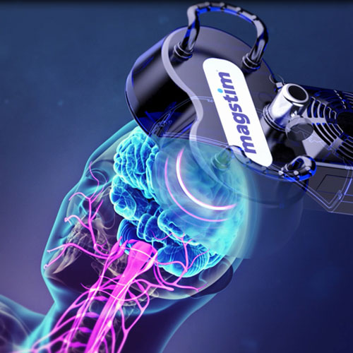
The biological effects of NIBS are essentially determined by two types of factors: extrinsic and intrinsic . On one hand, extrinsic factors are related to the amount of energy and to the pattern of current flow delivered to the brain. These include specific parameters that can be actively controlled by the operator, such as current intensity, stimulation frequency, number of pulses, number of sessions, coil design, electrode montage, etc. However, for the same dose of energy delivered, different intrinsic factors inherent to the stimulated subject contribute to the individuals biological outcome. For instance, the subjects pharmacological profile can affect the brains activation state and connectivity by modulating neuronal propensity to fire and undergo plastic phenomena. In patients with Parkinsons disease , this is particularly noteworthy, as changes in cortical excitability and neuroplasticity are critically influenced by dopamine bioavailability, and the institution of a dopaminergic therapy can influence the subsequent neurophysiologic and behavioral effects of stimulation .
Negative And Positive Effect In Rtms Trials
Although this meta-analysis shows a favourable effect of TMS on motor function in PD, a positive effect was not observed in every trial. One of the reasons may be the small sample size of these negative studies. In this scenario, the meta-analysis technique is a valuable method to combine the data from small studies in order to provide a conclusion based on an analysis with better power. However, two studies with relatively large sample sizes showed negative results. One explanation for this contradiction might be the interaction of antiparkinson drugs with TMS, as these studies assessed the motor UPDRS after the use of levodopa . This medication might mask the effects of TMS due to a ceiling effect. Therefore, assessment of patients in the off state may provide a more sensitive measure of the benefit of TMS. An alternative explanation is that the variability of the results stems from the wide range of TMS parameters and patient selection criteria used in these studies, that is, the optimal TMS parameters might vary depending on disease duration and severity. Although the meta-regression results failed to show that TMS parameters could significantly account for the variability across studies in motor improvement, the interaction term was not analysed because of lack of power for this type of test.
Studies Utilizing Theta Burst Stimulation
Unlike rTMS, the protocol of TBS is comparatively more consistent among studies . For all five studies utilizing continuous TBS , cTBS consists of three-pulse bursts at 50 Hz repeated every 200 ms for 40 s and was administered after levodopa intake.
In Koch’s study, they firstly applied single-session cTBS on 10 PD patients with LID over the cerebellum, and a 45-min reduction was observed . In this study, a 10-day course of cTBS was further conducted and induced persistent clinical beneficial effects up to 4 weeks . However, a later study applied a 5-day course of cTBS on 8 PD patients with LID over the cerebellum only reduced LID up to 45 min .
A study applied single-session cTBS over the inferior frontal cortex and MC on 8 PD patients with LID, respectively . Stimulation over the right IFC induced improvement of LID only up to 30 min, while stimulation over MC did not exhibit any change . Although efficacy duration was not mentioned, Ponza et al. also observed the beneficial effect of cTBS on LID symptoms after single-session stimulation over the right IFC . A recent study targeting cerebellum also displayed 60-min alleviation for LID after cTBS stimulation .
Like LF-rTMS, the short-term benefits of cTBS have been corroborated in several studies and are patient-tolerable. Although a remarkably longer after effect of cTBS than of LF-rTMS was exhibited only in one study, such prolonged effect did not replicate in other studies.
Read Also: Young Onset Parkinson’s Symptoms
Can You Treat Parkinsons Disease With Tms Therapy
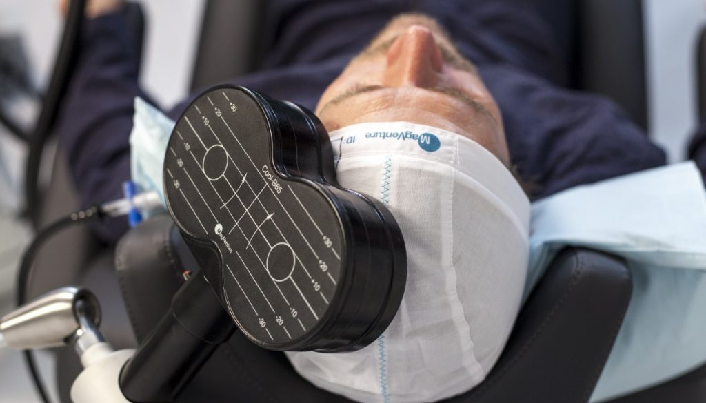
Can you treat Parkinsons disease with TMS therapy? Its an excellent question and most easily answered, yes, yes you can. However, it must be remembered that TMS, or transcranial magnetic stimulation therapy, is strictly thattherapy. Thus far, there is no cure for Parkinsons, but research and testing continue, and some strides have been made in the treatment of the disease. In the meantime, there are medications, physical therapy, and other treatments proven to help Parkinsons patients abate their symptoms and live longer and more comfortable lives.
In more recent years, TMS therapy for Parkinsons has been shown to help Parkinsons patients with controlling or diminishing such classic symptoms as freezing, tremors, and rigidity through delivering pulses to the parts of the brain related to these symptoms and their usual attendant movements. But TMS isnt a panacea, and it seems to have a greater effect on certain types of Parkinsons than on others. Regardless, results have been promising. With further study and testing, we may discover other ways TMS can help Parkinsons and other nerve and brain disorders like seizures, anxiety, depression, and more. Heres what we know so far and what we hope to learn more about in the future.
Smf Mediated Changes Are Consistent With L

To gain a better perspective whether long-lived changes could have been initiated through the proposed modulation of calcium channel activity by SMF, an independent method to alter Ca2+ flux was evaluated. Specifically, Bay K8644 and nifedipine, were used to alter Ca2+ flux in PC12 cells and A2AR mRNA levels were again evaluated. In this experiment, Bay K8644 increased A2AR mRNA levels while nifedipine treatment decreased transcription in essence Bay K8644 reproduced the effects of agonist }CGS21680 and nifedipine mimicked antagonist ZM241385 . To further strengthen the correlation between L-type Ca2+ channels, calcium flux, and A2AR transcription, we demonstrated that the increased levels of A2AR mRNA found in Bay 8644 treated cells could be reduced to levels found in control cells by concomitant exposure to SMF .
Effect of L-type Ca2+ channel activators and blockers on A2AR mRNA and protein levels in PC12 cells.
The L-type Ca2+ channel activator Bay K8644 increased A2AR mRNA levels in PC12 cells compared to untreated controls while the L-type Ca2+ blocker Nifedipine, as well as SMF exposure, decreased A2AR mRNA levels after 6.0 h of exposure . Increased A2AR mRNA resulting from exposure to Bay 8644 was reversed by concomitant exposure to SMF .
You May Like: Parkinson Disease Stem Cell Clinical Trials
The Nervous System And Parkinson’s Disease
for background information on this. So, one of the early major turnarounds I gained from using the PEMF device was that, if I put it on the back of my right shoulder , just as I sit down to dinner, and leave it there for an hour, then I found this actually prevented much of the subsequent symptomatic shut down and medication ineffectiveness due to digestive impacts.
What Can Tms Help And When Is It Used
Can you treat Parkinsons disease with TMS therapy? Research and testing show that TMS therapy is best suited for fighting the symptoms of akinetic-rigid Parkinsons as opposed to tremor-dominant Parkinsons. Why? Its not yet entirely clear, but the prospects are promising. As for when it should be used, that depends on a doctor or neurologists recommendation, and usually when they determine that medication and physical therapy by themselves arent helping control the symptoms.
Other things that make a patient a candidate for TMS therapy for Parkinsons include symptoms severely cutting into a patients ability to function and enjoy life, as well as an increase in involuntary movements and tremors. If the amount of medication a patient takes each day has reached four or more doses, thats another red flag for the use of TMS treatment. As a side note, TMS isnt used alone, and is most often combined with other therapies and treatments. Transcranial magnetic stimulation is usually combined with aerobic activity to help increase strength as well as to test the connection between the brain and the other parts of the body.
You May Like: Gender Differences In Parkinson’s Disease
Pemf Therapy For Parkinsons Treatment
Parkinsons disease is a brain disease . Parkinsons disease symptoms can be different, and for each person, Parkinsons disease acts differently. The disease is characterized by a progressive motor disorder that severely affects the daily life of the patients.
Recently researcher Moniek Munneke of Radboudumc developed a procedure known as Electromyography charted the connection between the brain and muscles using magnetic stimulation. When a magnetic field is generated above the brain region that drives movements, this leads to involuntary contraction of the muscles on the other side of the body than where the stimulation was given. The effect of magnet stimulation on the muscle tension can be measured by electromyography . Moniek Munneke: If we can move the paralysed muscle through the external stimulus, this means that the nerve pathways between the brain and the muscles are still intact. With this information, we can finally help patients with an expectation of recovery. It is imperative in this technique where you stick the electrodes to the skin for the EMG measurement. Through our research, we now know enough to test this method for use in Parkinsons recovery.
Moniek then went on to do further research covered in her dissertation, MEASURING AND MODULATING THE BRAIN WITH NON-INVASIVE STIMULATION, an excellent read for doctors and PEMF device developers.
J NeurolBehav Brain FunctClin Neurol NeurosurgFront NeurosciNeuroscientistTransl NeurodegenerNeurol Sci
Repetitive Transcranial Magnetic Stimulation In Parkinsons Disease
Two mechanisms have been proposed to explain how cortically directed rTMS may improve PD symptoms: rTMS induces brain network changes and positively affects the BG function rTMS directed to cortical sites compensates for PD-associated abnormal changes in cortical function . Indeed, in support of the former mechanism, rTMS might modulate cortical areas, such as the prefrontal cortex and primary motor cortex, which are substantially connected to both the striatum and STN via glutamatergic projection, and thus indirectly modulate the release of dopamine in the BG . Several TMS/functional imaging studies have demonstrated the effects of rTMS on BG and an increase in dopamine in the BG after rTMS applied to the frontal lobe .
Cognitive dysfunction is often seen in PD patients with major depression and its neural basis could be the functional failure of the frontostriatal circuit . Ten days of rTMS in the frontal cortex can effectively alleviate PD-associated depression as shown by an open trial reporting a significant decrease in the Hamilton Depression Rating Scale scores . A further double blind, sham stimulation-controlled, randomized study, involving 42 idiopathic PD patients affected by major or minor depression undergoing rTMS for 10 days, evidenced a mean decrease in HDRS and Beck depression inventory after therapy .
Read Also: Agent Orange And Parkinson Like Symptoms
Smf Impinges Upon Mapk Pathways And Impacts Pc12 Cell Proliferation
Stimulation of PC12 cells with }CGS21680 increases the phosphorylation of p44/42 MAPK via cAMP-mediated signaling , . This prior observation, together with known links between NO production and phosphorylation of p44/42 MAPK , prompted us to test whether SMF and the A2AR modulators }CGS21680 and ZM241385 also affected p44/42 MAPK. Accordingly, we first investigated whether }CGS21680 increased the phosphorylation of p44/42 MAPK and observed an increase by Western blot analysis after 30 min of exposure that was consistent with enhanced proliferation observed in the agonist-treated cells . By contrast, pretreatment of the cells with the ZM241385 or co-treatment with SMF reversed }CGS21680-induced p44/42 MAPK phosphorylation resulting in levels lower than observed in untreated control cells the accelerated proliferation observed in }CGS21680 treated cells also was not seen under these condition . In these experiments, SMF by itself also reduced levels of phospho-p44/42 MAPK and proliferation.
Effect of }CGS21680, ZM241385, and SMF on p44/42 MAPK phosphorylation and proliferation in PC12 cells.
Electromagnetism In And Around Us
On earth, we are surrounded by electromagnetic fields, which are invisible to the human eye. To begin with, the Earth itself is a magnet with a north and a south pole. The magnetic field around the earth is essential for all life on earth. This protects us, among other things, from dangerous solar radiation. Birds, fish and certain insects are then dependent on the earth magnetic field to orient themselves.
In addition to this natural magnetic field, electromagnetic fields are also produced by all kinds of electrical appliances, such as TV, computer, microwave, electrical wiring, telecommunications.
All of us are exposed to different electrical and electromagnetic fields daily. It must have an effect on us, as even in our body, small electric and electromagnetic currents are generated and these can receive interference from EMF radiation.
As a result of the ever-increasing environmental pollution and electrosmog, our bodys magnetic equilibrium is always under high pressure and ultimately obstructs equilibrium. In this way, the proper functioning of our cellular metabolism is also getting more and more affected1. It could be one of the Parkinsons disease causes as almost every disease can be attributed to cellular dysfunction.
1. DAngelo C, Costantini E, Kamal M, Reale M. Experimental model for ELF-EMF exposure: Concern for human health. Saudi J Biol Sci. 2014 22:75-84. PMC
Read Also: Does Parkinson Cause Muscle Spasms
Master Pd: Magnetic Stimulation For The Treatment Of Motor And Mood Symptoms Of Parkinson’s Disease: A Four
Objective/Rationale:To determine the efficacy and duration of benefit of noninvasive brain stimulation with repetitive transcranial magnetic stimulation to modulate brain activity in order to improve motor and mood symptoms in Parkinson’s disease. Project Description: At four leading centers in North America we will study a total of 160 patients with idiopathic PD. Patients will have significant motor problems despite treatment with medications and meet criteria for a depressive disorder.
Whats The Best Way For Me To Diagnose Parkinsons

If you have any of these symptoms or know someone who does, its best to see your doctor so they can take blood tests and rule out other conditions that may be causing your symptoms. They might refer you to a specialist where they will take further tests such as MRI, CT, ultrasound of the brain, and PET scans. However, Imaging tests arent particularly accurate for diagnosing PD.
A movement disorder specialist is best suited to diagnose Parkinsons disease. If you think you or someone you love might have it, the Parkinsons Foundation website has a provider search resource.
Read Also: Atrial Fibrillation And Parkinson’s
Exposure Of Cells To Smf
A problem hindering the acceptance of magnetic therapy has been that many studies have used inadequately defined treatment devices leading to difficulties reproducing experimental conditions from laboratory to laboratory . Thus, in a previous publication we carefully described our magnetic exposure conditions . Neodymium magnets are arranged in this device to provide a unidirectional field that varies between 0.23 and 0.28 T over several centimeters . Consequently, the shallow nature of this gradient ensures that individual cells are essentially exposed to uniform fields and are not subject to spatial gradient effects observed in cells exposed to fields that varied by â¥20 mT/mm â. Moreover, no dose-dependency was observed between 0.23 and 0.28 T for the endpoints tested in this report . Untreated control cells were maintained in a separate tissue culture incubator where the ambient magnetic field was measured at â¼52 mT, which is essentially identical to the 52,359 nT field reported for a latitude of 39° 19â² 35â³ and a longitude of â76° 36â² 17â³ by the National Geophysical Data Center). Finally, we have tested whether orientation of the imposed field superimposed on â or opposing â the ambient geomagnetic field affects the endpoints being studied field orientation was found to not have an effect .
What Can It Treat
Currently, TMS is only FDA-approved to treat Major Depression Disorder. However, other disorders are suspected to be respondent to TMS as well. These include bipolar disorder, attention-deficit/hyperactivity disorder, panic disorder, obsessive compulsive disorder, schizophrenia, ALS, and Parkinsonâs.
You May Like: What Can Help Parkinson’s Disease
Magnetic Stimulation For Parkinson Disease
| The safety and scientific validity of this study is the responsibility of the study sponsor and investigators. Listing a study does not mean it has been evaluated by the U.S. Federal Government. Read our disclaimer for details. |
| First Posted : January 10, 2002Last Update Posted : August 18, 2006 |
- Study Details
| Procedure: Prefrontal transcranial magnetic brain stimulation | Phase 1 |
Inclusion Criteria:
- Have a diagnosis of idiopathic Parkinson’s Disease and meet DSM-IV criteria for Major Depressive Episode, severe, with or without psychotic features, or for Mood Disorder secondary to PD with major depression-like episode.
- Have demonstrated an inadequate clinical response to at least one antidepressant medication in adequate dosage for at least six weeks, or an adverse event requiring discontinuation.
Information from the National Library of Medicine
To learn more about this study, you or your doctor may contact the study research staff using the contact information provided by the sponsor.
Please refer to this study by its ClinicalTrials.gov identifier : NCT00029276
Smf Exposure Increases Intracellular Camp Levels
Levels of cAMP are another parameter relevant to PD that can be interrogated in PC12 cells this ubiquitous second messenger is linked to Ca2+ through a complex sequence of events mediated by A2AR and Gαs proteins . To evaluate connections between cAMP and A2AR in our experiments, we analyzed cAMP levels in agonist and antagonist treated cells and found a modest increase in the former and a more substantial decrease in the latter . In these experiments SMF decreased cAMP levels, again showing that magnetic exposure can functionally reproduce the cellular effects of an A2AR antagonist.
cAMP levels in SMF, }CGS21680, and ZM241385 treated PC12 cells.
Cells were exposed to each condition, harvested, lysed, and assayed for cAMP levels. Each test condition treatment condition varied from untreated control cells with p< 0.05 for n=3 independent experiments.
You May Like: Classification Of Parkinson’s Disease