Substantia Nigra And Parkinson’s Disease: A Brief History Of Their Long And Intimate Relationship
Published online by Cambridge University Press: 02 December 2014
- Department of Psychiatry and Neuroscience, Laval University School of Medicine, Quebec City, Quebec, Canada
- André Parent*
- Department of Psychiatry and Neuroscience, Laval University School of Medicine, Quebec City, Quebec, Canada
- *
Motor Circuit In Parkinson Disease
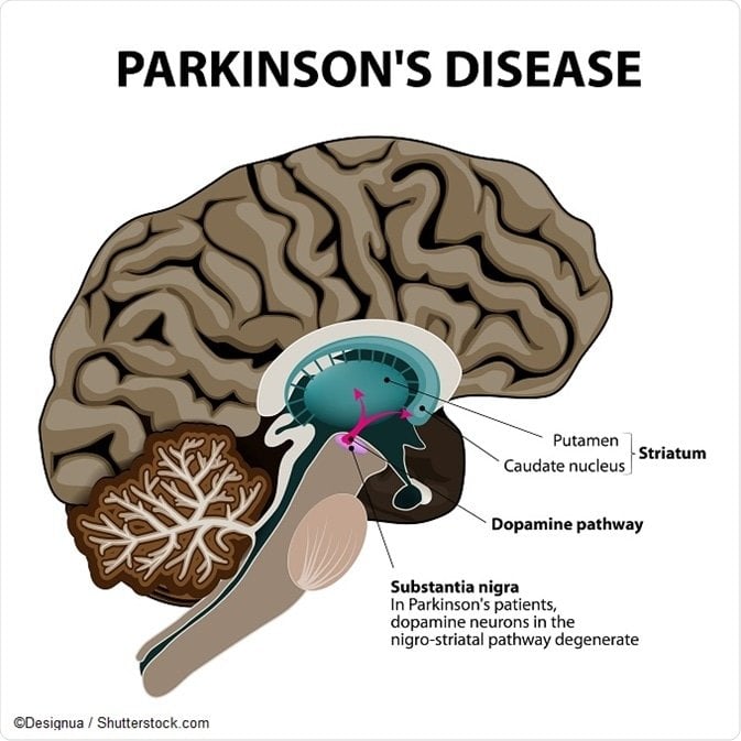
The basal ganglia motor circuit modulates the cortical output necessary for normal movement .
Signals from the cerebral cortex are processed through the basal ganglia-thalamocortical motor circuit and return to the same area via a feedback pathway. Output from the motor circuit is directed through the internal segment of the globus pallidus and the substantia nigra pars reticulata . This inhibitory output is directed to the thalamocortical pathway and suppresses movement.
Two pathways exist within the basal ganglia circuit, the direct and indirect pathways, as follows:
-
In the direct pathway, outflow from the striatum directly inhibits the GPi and SNr striatal neurons containing D1 receptors constitute the direct pathway and project to the GPi/SNr
-
The indirect pathway contains inhibitory connections between the striatum and the external segment of the globus pallidus and between the GPe and the subthalamic nucleus striatal neurons with D2 receptors are part of the indirect pathway and project to the GPe
The STN exerts an excitatory influence on the GPi and SNr. The GPi/SNr sends inhibitory output to the ventral lateral nucleus of the thalamus. Dopamine is released from nigrostriatal neurons to activate the direct pathway and inhibit the indirect pathway. In Parkinson disease, decreased striatal dopamine causes increased inhibitory output from the GPi/SNr via both the direct and indirect pathways .
Contribution Of The Substantia Nigra
Damage to the SN pars compacta not only leads to a depletion of dopamine in the nigrostriatal pathway but may also cause dopamine deficiency in the nigro-subthalamic and nigro-pallidal pathways. In human brains, the SN pars compacta send dopaminergic projections to the subthalamic nucleus and the internal and external segments of globus pallidus in addition to the striatum. In PD patients, dopamine levels in the subthalamic nucleus and globus pallidus decrease remarkably, similar to that in the striatum . Dopamine depletion might lead to the observed weakened functional connectivity between the SN and basal ganglia.
In 6-hydroxydopamineinduced rat models of PD, the firing rate of subthalamic neurons starts to increase one day after the unilateral lesion of the SN pars compacta and remains significantly higher than that in normal controls 2 weeks after the lesion, which is accompanied by irregular and bursty firing patterns . In contrast, the firing rate of pallidal neurons decreases prominently 3 weeks after the lesion of the striatum but not after the lesion of the SN pars compacta . Similar changes in neuronal activity might underlie the observed subthalamic hyperactivation and pallidal hypoactivation. The subthalamic hyperactivation and caudate hypoactivation might be consequences of SN damage, whereas the pallidal hypoactivation is likely a consequence of caudate hypoactivation.
Read Also: Judy Woodruff Parkinson’s
The Nervous System & Dopamine
To understand Parkinson’s, it is helpful to understand how neurons work and how PD affects the brain .
Nerve cells, or neurons, are responsible for sending and receiving nerve impulses or messages between the body and the brain. Try to picture electrical wiring in your home. An electrical circuit is made up of numerous wires connected in such a way that when a light switch is turned on, a light bulb will beam. Similarly, a neuron that is excited will transmit its energy to neurons that are next to it.
Neurons have a cell body with branching arms, called dendrites, which act like antennae and pick up messages. Axons carry messages away from the cell body. Impulses travel from neuron to neuron, from the axon of one cell to the dendrites of another, by crossing over a tiny gap between the two nerve cells called a synapse. Chemical messengers called neurotransmitters allow the electrical impulse to cross the gap.
Neurons talk to each other in the following manner :
Brainbehavior And Structurefunction Relationships

First, the ordering-related accuracy cost correlated with the ordering-related basal ganglia activation in both groups but in different manners . In HCs the stepwise regression model = 4.59, p = 0.04, R2 = 0.18) included the ordering-related subthalamic activation but removed the ordering-related caudate activation and total SN-caudate/subthalamic functional connectivity . In PD patients, the stepwise regression model = 11.40, p = 0.003, R2 = 0.32) included the ordering-related caudate activation but removed the ordering-related subthalamic activation and total SN-caudate/subthalamic functional connectivity . HCs with greater ordering-related subthalamic activation and PD patients with greater ordering-related caudate activation exhibited smaller accuracy costs for sequence manipulation.
To confirm the SN effect on the ordering-related caudate activation, we conducted an SPM12 whole-brain one-sample t test in PD patients with the total SN area as a covariate . C presents the SN effect in the left caudate nucleus . There was no significant cluster in the subthalamic nucleus or globus pallidus.
Don’t Miss: Does Vitamin B12 Help Parkinson’s
Group Differences In Task Accuracy
D presents task accuracy of the digit-ordering task in each group. We examined group differences in accuracy using an ANOVA with two factors, Group and Trial Type . Main effects of Group = 7.93, p = 0.007, 2 = 0.12) and Trial type were found = 3.57, p = 0.06, 2 = 0.06), but no interaction . It replicated previous findings that participants tended to be less accurate in reorder and recall than pure recall trials , and that accuracy of PD patients was less accurate than that of HCs.
A Robust Method For The Detection Of Small Changes In Relaxation Parameters And Free Water Content In The Vicinity Of The Substantia Nigra In Parkinsons Disease Patients
-
Roles Conceptualization, Data curation, Formal analysis, Investigation, Methodology, Software, Validation, Visualization, Writing original draft, Writing review & editing
Affiliation Institute of Neuroscience and Medicine 4 , Forschungszentrum Jülich GmbH, Jülich, Germany
-
Roles Conceptualization, Formal analysis, Investigation, Supervision, Validation, Writing original draft, Writing review & editing
Affiliation Institute of Neuroscience and Medicine 4 , Forschungszentrum Jülich GmbH, Jülich, Germany
-
Roles Conceptualization, Formal analysis, Investigation, Methodology, Writing original draft, Writing review & editing
Affiliation Institute of Neuroscience and Medicine 4 , Forschungszentrum Jülich GmbH, Jülich, Germany
-
Roles Conceptualization, Resources, Validation, Writing review & editing
Affiliation Department of Nuclear Medicine and PET Center, Aarhus University, Aarhus, Denmark
-
Roles Resources, Supervision, Writing review & editing
Affiliation Department of Nuclear Medicine and PET Center, Aarhus University, Aarhus, Denmark
Don’t Miss: Parkinson Bicycle Cleveland Clinic
Quality Control And Data Correction
Quality control and data correction for single-cell samples were based on the number of detected genes, the number of detected molecules, and the percentage of mitochondrial, ribosomal and hemoglobin genes from each single-cell sample. Individualized filtering plans were applied to different datasets based on their unique features . In detail, for dataset , samples with less than 1000 genes, more than 3000 gene, less than 200 molecules, more than 1% mitochondria genes and more than 2% ribosomal genes were removed. For dataset , samples with less than 600 genes, more than 2000 genes, less than 200 molecules, more than 0.5% mitochondrial genes and more than 2% genes were removed. For dataset , single-cell samples with less than 1000 genes, more than 6000 genes, less than 200 molecules, more than 0.05% ribosomal genes were removed. The remaining data in the three datasets were later used to produce a combined dataset.
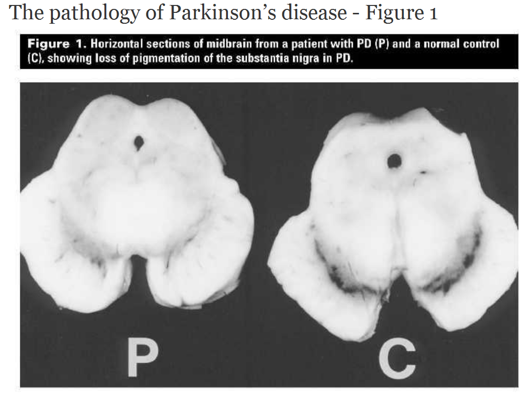
Heterogeneous Patterns Of Cell Loss In The Substantia Nigra In Parkinson’s Disease
There were highly significant differences in the extent of cell loss in the different subgroups of the nigral complex. Loss was higher in the substantia nigra pars compacta than in the substantia nigra pars dorsalis , and there was a small loss , which did not reach statistical significance, in the substantia nigra par lateralis .
The calbindin-based definition of compartments within the substantia nigra pars compacta demonstrated further heterogeneity in neurodegenerative patterns. First, as shown in Table 3, the cell loss was significantly greater in the nigrosomes than in the matrix . This was true for each rostrocaudal level for all nigrosomes, excepting only the caudal nigral levels in patient P974, in which cell loss was nearly total in both nigrosomes and matrix . Secondly, there were clear differences in the loss of dopamine-containing neurons in the different nigrosomes . The mean cell loss was maximal in nigrosome 1 . The few TH-positive neurons that did survive in nigrosome 1 did not appear to have characteristic locations . Nigrosome 2 and nigrosome 4 were the next most affected, and nigrosome 3 and nigrosome 5 were considerably less affected. The degree of loss of dopamine-containing neurons in the different nigrosomes was strictly ordered: nigrosome 1 > nigrosome 2 > nigrosome 4 > nigrosome 3 > nigrosome 5.
Recommended Reading: Judy Woodruff Health Problems
Normalization Integration Dimensionality Reduction Clustering And Visualization And Cluster Annotation
After quality control, Seurat R package v4.0.2 was used to process the data. Sctransform , which enables the recovery of sharper biological distinction, was used for normalization. Then, Harmony, an integration algorithm, was used to integrate the above-mentioned three datasets and perform the dimensionality reduction . Clustering and visualization of the integrated dataset were realized by Uniform Approximation and Projection method at a resolution of 0.05 and a dimension of 20. Canonical marker genes for neuronal and non-neuronal cells were used to annotate each cell cluster.
Dopamine Depletion Decreases Excitability Of Gabaergic Neurons In The Snr
Spontaneous firing of GABAergic neurons, in the presence of blockers for synaptic transmission, was affected by dopamine depletion, probably due to changes in intrinsic neuronal properties. The decrease in the firing rate of dopamine depleted rats observed here is consistent with previous work on the SNc-SNr pathway which suggests the existence of a pathway where dopamine released from dendrites of SNc neurons binds to D1 and D5 receptors on GABA neurons in the SNr. The coactivation of the two receptors enhances the activity of TRPC3 channels, thus leading to depolarization. Inhibition of these receptors led to a reduction in firing rate and an increase in the irregularity of ISI, which may imply that in dopamine depleted rats, there was no dendritic release of dopamine, no TRPC3 enhancement and thus a reduction in the spontaneous firing rate in comparison to the naïve rats. These findings are also in line with recent work showing a reduction in firing rate, under two conditions namely, the blockage of D1 and D2 receptors and experimental Parkinsonism in juvenile mice . We also identified an increase in irregularity, as did Zhou et al. . In contrast to in vivo studies that have reported an increase in the bursting activity of SNr neurons , we rarely encountered bursting activity, which may be due to the use of blockers for glutamatergic inputs in our experiments .
You May Like: Weighted Silverware
Are Alterations In The Axonal And Synaptic Mitochondrial Populations Correlated With Respiratory Deficiency In The Soma
Although in human tissue it is not possible to compare cell body changes to synaptic/axonal changes within the same neuron, a measure of respiratory chain deficiency was made within the soma of SN neurons for most of the cases included in this study . Intensity measurements were made for immunoreactivity for NDUFB8, COXI and porin within TH positive cell bodies within the SN. A reduction in expression of either of these proteins was defined as an intensity value that had a z score of less than 1, normal neurons had a score of between 1 and 1, while an increase was defined as having a score over 1. These parameters were set based on the control data.
There was an increase in the percentage of cell bodies showing reduced expression of both complex I and complex IV in PD cases compared to other groups, and in DLB cases there was an increase in complex IV deficient neurons, however neither of these changes was significant due to the variability between cases . In addition to measuring deficiency within the neurons of the SN, mitochondrial density was also quantified. Using the signal intensity for porin immunoreactivity we categorised each neuron based on the z score for porin. We again found that there was no significant difference between the mitochondrial mass in cell bodies between any of our patient groups .
Data And Statistical Analysis

All off-line analyses were carried out using Matlab R2013a and IgorPro 6.0 on a personal computer. All results for each experiment were pooled and displayed as the mean ± SEM unless mentioned otherwise. We calculated spontaneous firing rates and coefficients of variation from stable recordings lasting 10 s and recorded evoked IPSPs and miniature IPSCs at a holding potential of â60 mV. We calculated evoked IPSP latency as the time from the end of the stimulation to the onset of the current deflection. We detected mIPSCs with an Offline Sorter program . We analyzed confocal images with ImageJ in conjugation with the Synapse Counter plug-in to identify GABAergic puncta. In all figures comparing between naïve and dopamine depleted animals, we used the MannâWhitney U-test . The curves in Figures 3D, 4B were compared using bootstrap. The bootstrap method is a resampling technique used to estimate mean or standard deviation on a population by sampling a dataset with replacement . We used it to estimate whether the difference between the curves fitted to the data in these figures belonged to the same distribution. The data inFigure 6 was compared using the permutation test.
Recommended Reading: On Off Phenomenon
Mapping And Quantitative Analysis Of Dopamine
Dopamine-containing neurons in the midbrain were identified as TH-positive neurons. Charts of the distributions of dopamine-containing neurons were constructed from every TH-stained section by plotting TH-positive neurons with a computer-assisted image analysis device . Five dopaminergic cell groups were identified in the midbrain , and the substantia nigra was subdivided into the substantia nigra pars compacta, formed by the matrix and five nigrosomes, the substantia nigra pars dorsalis and the substantia nigra pars lateralis, according to the patterns of calbindin immunostaining observed. This plan of subdivision is described in detail in the accompanying paper .
The total numbers of dopamine-containing neurons in each cell group were calculated for each patient by integrating values for individual sections over the entire length of the midbrain. The percentage of cell loss in Parkinson’s disease was calculated from these values by comparing means for parkinsonian and control midbrains. Split-cell counting errors were corrected by applying the formula of Abercrombie . The correction factor was 0.61 no significant differences were found between the sizes of neuronal nuclei in control and Parkinson’s disease brains.
Contribution Of D2/3 Receptor Agonists
This study is consistent with previous findings that dopamine plays a role in sequential working memory . However, the exact dopaminergic mechanism is an open question. In visuospatial working memory, it is proposed that D1 receptors mediate the robust maintenance, whereas D2 receptors mediate the dynamic updating . However, evidence for differential roles of D1 versus D2 receptors in sequential working memory is insufficient and conflicting. For example, observed improved sequencing performance in de novo PD patients after a 4-month monotherapy of levodopa or bromocriptine . In contrast, observed improved sequencing performance in healthy adults after a single dose of 400 mg sulpiride versus a placebo. In addition, observed an interaction between sequential working memory and a 2-month treatment of piribedil in healthy adults after the treatment, participants with a higher capacity showed an improvement, whereas those with a lower capacity showed impairment in verbal fluency.
Recommended Reading: Pfnca Wellness Programs
Differential Vulnerability Of The Nigral Compartments Nigrosomes And Matrix In Parkinson’s Disease
A major finding of this study is that, in every parkinsonian midbrain, dopamine-containing neurons in nigrosomes were more affected than dopamine-containing neurons of the matrix. Based on the subdivisions defined by calbindin patterns in neurologically normal midbrains , dopamine-containing neurons of the substantia nigra pars compacta can be divided into two types: sparsely distributed neurons included in a calbindin-rich matrix compartment, and densely packed neurons in five calbindin-poor nigrosomes. Our quantitative estimates in control midbrains suggest that the population of the matrix neurons is normally ~1.4 times larger than the population of neurons in the nigrosomes . The consistent difference in cell loss in matrix and nigrosomes in all five parkinsonian patients, found at all levels in the substantia nigra pars compacta containing surviving neurons, suggests that differential nigrosome/matrix vulnerability may be a basic attribute of the disease process in Parkinson’s disease.
What Are The Causes
The cause of Parkinson’s is largely unknown. Scientists are currently investigating the role that genetics, environmental factors, and the natural process of aging have on cell death and PD.
There are also secondary forms of PD that are caused by medications such as haloperidol , reserpine , and metoclopramide .
Don’t Miss: Cleveland Clinic Parkinson’s Bicycle Study 2017
Other Causes Of Parkinsonism
“Parkinsonism” is the umbrella term used to describe the symptoms of tremors, muscle rigidity and slowness of movement.
Parkinson’s disease is the most common type of parkinsonism, but there are also some rarer types where a specific cause can be identified.
These include parkinsonism caused by:
- medication where symptoms develop after taking certain medications, such as some types of antipsychotic medication, and usually improve once the medication is stopped
- other progressive brain conditions such as progressive supranuclear palsy, multiple systems atrophy and corticobasal degeneration
- cerebrovascular disease where a series of small strokes cause several parts of the brain to die
You can read more about parkinsonism on the Parkinson’s UK website.
Page last reviewed: 30 April 2019 Next review due: 30 April 2022
Group Differences In Substantia Nigra Areas
E presents SN areas in each group. We examined group differences in the SN area using an ANOVA with two factors, Group and Side . Main effects of Group = 9.87, p = 0.003, 2 = 0.15) and Side = 66.39, p< 0.001, 2 = 0.55), and an interaction between Group and Side were found = 14.11, p< 0.001, 2 = 0.20). The left SN was larger than the right SN in both groups. PD patients showed smaller SNs than HCs, especially on the left side. In PD patients, the total SN area correlated with the UPDRS III nontremor score . PD patients with smaller SNs showed more severe nontremor symptoms . There was no correlation between the SN area and UPDRS III tremor score .
Read Also: Sam Waterston Tremor