Integrated Evidence For Paraquat Hazard Identification
For proper categorization of paraquat in one of the five categories of hazard , evidence coming from human and animal studies will be integrated with mechanistic data that may be relevant to support biological plausibility and increase or decrease the hazard classification.
Among the factors that can support biological plausibility and increase the hazard classification, the magnitude of the effect, the doseresponse gradient, and the direct and indirect consistencies between outcomes from studies with different biological levels will be considered. On the contrary, hazard classification may be reduced by the identification of risk of bias, unexplained inconsistencies between studies with related outcomes, non-relevance of the paraquat mechanism of toxicity to humans and dose levels not relevant to real human exposure.
Also Check: Similar To Parkinsons
Analysis Of Dopamine Dopac And Hva
Six mice of each group were anaesthetized with isoflurane inhalation. The striatum was collected and then was homogenized in ice-cold 0.1M HClO4 containing 0.01% EDTA. After centrifuged , the concentration of dopamine and its metabolites homovanillic acid and 3,4-dihydroxyphenylacetic acid in supernatant were assayed by high-performance liquid chromatography with fluorescence. Concentrations of dopamine, HVA, and DOPAC were expressed as g/g tissue weight.
Ultrastructure Detection By Transmission Electron Microscopy
After the rats were anaesthetized and perfused, the SN was isolated and cut into approximately 1-mm cubes. The samples were fixed with a solution of 2.5% glutaraldehyde with 2% paraformaldehyde, and then exposed to 1% osmium tetroxide. After several subsequent washes, the tissue samples were dehydrated with gradient alcohol, embedded in Epon and finally polymerized. Regions of interest were identified and the ultrathin sections were spread on the grids. After the sections were contrasted with uranyl acetate and lead citrate, observations were performed using a transmission electron microscopy .
You May Like: Cleveland Clinic Parkinson’s Bicycle Study 2017
Drug And Chemical Agents
Danshensu was from Shandong Luye Pharmaceutical Co., Ltd. . Rotenone, dimethylsulfoxide , streptomycin/penicillin, dopamine, DCFH-DA, and GSH were purchased from Sigma Chemical Company . Dulbeccos modified Eagles medium was from Gibco Company . Fetal bovine serum was provided by Zhejiang Tianhang Biotechnology Co., Ltd. .
Rotenone Toxicity Is Caused By Complex I Inhibition

Rotenone is believed to be a specific inhibitor of complex I of the mitochondrial ETC. To determine whether rotenone toxicity depended on interaction with complex I, we analyzed toxicity in SK-N-MC cells expressing the rotenone-insensitive single-subunit NADH dehydrogenase of Saccharomyces cerevisiae , which acts as a replacement for the entire complex I in mammalian cells . Malate-glutamate-driven mitochondrial respiration was increased twofold to threefold in NDI-1-transduced cells. Oxygen utilization in NDI1-transduced cells, using complex I substrates , was rotenone-insensitive, but remained sensitive to the complex III inhibitor antimycin A, indicating that NDI1 integrated into the mitochondrial ETC and donated electrons appropriately to complex III .
Recommended Reading: Parkinson Silverware
Animals And Ethics Approval
In this study, adult male Swiss albino mice were used. Mice were purchased from the M. Rashed Company for experimental animals and kept under clean laboratory settings and a normal lightdark cycle. Water and food were allowed ad libitum. Male Swiss albino mice were selected as they have been frequently used in previous models of parkinsonism . Experimental protocols and animal handling procedures were officially approved by the research ethics committee , Faculty of Pharmacy, Suez Canal University, in compliance with the National Institute of Health Guide for the Care and Use of Laboratory Animals .
Modelling Pd In Rodents And Simple Organisms
Impaired mitochondrial function is a predominant feature in cases of Parkinsons disease due to genetic or environmental modifications, which result in mitochondrial stress. This may directly compromise the neuron or cause alterations in neurotransmitter release, resulting in post-synaptic damage. A pro-inflammatory component is increasingly considered to be important in the pathogenesis of several neurodegenerative conditions including PD, and pro-inflammatory activation of glial cells may be pivotal in disease onset.,
Although many of the genes involved in PD have been identified, their interactions are still unclear. Mutations of -synuclein or duplications of -synuclein are linked to familial PD and -synuclein is a component of Lewy bodies. -synuclein tends to form intracellular fibrils and aggregates, particularly after oxidative stress. Loss of an ubiquitin ligase, Parkin, is responsible for autosomal recessive juvenile parkinsonism. Finally, mutations in genes coding for UCH-L1 and Pink-1 are linked to autosomal recessive PD, whereas mutations in LRRK2 are linked with autosomal dominant PD. Recent work has shown that mutations in the mitochondrial protease HtrA2 are also linked to PD downstream of the kinase, Pink 1. Mutations in Parkin are recognized as the most common cause of familial parkinsonism and may be involved in sporadic PD. Parkin seems to work as a broad-spectrum neuroprotectant, the efficacy of which decreases with ageing.
Dont Miss: Zhichan Capsule
Read Also: Parkinson’s Double Vision
Overview Of The Rodent Rotenone Models Of Parkinson’s Disease And Their Ability To Reproduce The Features Of Clinical Disease
The rotenone model largely only gained attention following the seminal paper by the Greenamyre group, which demonstrated for the first time that rotenone administered systemically to rats can reproduce the hallmarks of PD . Degeneration induced in the rodent rotenone model reproduces the degeneration in clinical PD , with damage occurring in the dopaminergic nerve terminals of the striatum and cell bodies in the SN . Furthermore,
What Is Parkinsons Disease
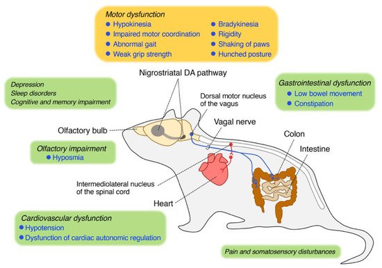
Parkinsons disease is a movement disorder in which the nerve cells that transmit messages from your brain and spinal cord to your muscles become damaged. That damage results in progressively worsening motor function and symptoms such as:
- Tremors in your arms, legs, hands, and face
- Slowed bodily movements
- Difficulties with balance, coordination, and speaking
- Stiffness in your arms and legs
- Nonmotor symptoms like depression, fatigue, and cognitive impairment
These symptoms are caused by damage and destruction to dopamine neurons in a persons brain and spinal cord a process called neurodegeneration. However, its unclear exactly why this cell damage happens in people with PD.
Also Check: Judy Woodruff Parkinson’s
Effect Of Danshensu On Rotenone
As shown in Fig. a, when exposed to Danshensu at concentrations of 10M or lower, the viability of SH-SY5Y cells had no significant difference as compared with the control cells. When exposed to rotenone for 24h, a significant decrease of cell viability was observed with 10nM and more significant toxicity was seen in 200nM and 400nM . Consequently, 100nM of rotenone was used to induce cytotoxicity in the subsequent experiments. SH-SY5Y cells were pretreated with Danshensu for 2h and then challenged with rotenone at concentration of 100nM for 24h. As shown in Fig. c, Danshensu significantly attenuated the cell toxicity induced by rotenone when compared with the rotenone group.
Fig. 1
Exposure To Paraquat: What Are The Risks
In 1997, the Environmental Protection Agency announced that exposure to Paraquat primarily happens during the application and post-application of the herbicide.
And even though only licensed applicators are given access to the chemical, exposure is still possible for individuals who reside near farms where Paraquat is used.
In which case, Paraquat poisoning is possible.
Recommended Reading: Zhichan Capsule
Assessments Of Mitobiogenesis In Rotenone
The mitochondrial mass was measured using the fluorescent dye NAO . NAO was added to SH-SY5Y cells to achieve a final concentration of 10M for 10min at 37°C after the cells were treated. The fluorescence intensity was detected and analysed with a Cellomics ArrayScan VTI HCS Reader with the Morphology Explorer BioApplication software. The mitochondrial mass was quantified by the value of the average fluorescent intensity of NAO.
Quantification of mtDNA was carried out using real-time qPCR. Total DNA was extracted using a DNAiso Reagent Kit , followed by qPCR using SYBR® Fast qPCR Mix . The relative mtDNA copy number was calculated as the ratio of mtDNA/nDNA. The primer sequences are listed in Supplementary Table .
For reverse transcription -qPCR, total RNA was extracted using the TRIzol reagent . RT-qPCR was performed using the PrimeScript RT Reagent Kit and SYBR® Fast qPCR Mix in accordance with the manual guide. -ACTIN mRNA was used as the internal control. The primer sequences are listed in Supplementary Table .
Signs Of Paraquat Poisoning

After a person ingests or inhales a toxic amount of Paraquat, he or she is likely to have swelling and pain in the mouth and in the throat.
The herbicide causes immediate damage through direct contact.
What follows are gastrointestinal symptoms, such as:
- nausea
- abdominal pain
- diarrhea that may be bloody
These gastrointestinal problems tend to be severe, and may result in dehydration, low blood pressure, and electrolyte abnormalities.
Even the ingestion of small to medium amounts of Paraquat may cause a person to experience heart failure, kidney failure, liver failure, lung scarring, respiratory failure, and failure of multiple organs within several days to several weeks.
On the other hand, the ingestion of large amounts of Paraquat will result in severe symptoms that may occur within several hours to several days, including:
- confusion
- liver failure
- respiratory failure which could possibly lead to death
Despite these dangers related to the use of the toxic chemical, Paraquat use in the US is still on the rise.
In fact, according to a 2016 report from the National Water-Quality Assessment project, usage of the herbicide in the country has doubled between 2006 and 2016.
That increase in use of Paraquat means that there will also be an increase in exposure to Paraquat, as well as in the harms associated with the chemical.
The bad news?
Recommended Reading: On-off Phenomenon
Determination Of Striatal Malondialdehyde Reduced Glutathione Hemoxygenase
Spectrophotometric measurement of reduced glutathione and malondialdehyde was done using kits obtained from Biodiagnostics . Using enzyme-linked immunosorbent assay kits, striatal levels of hemoxygenase-1 and dopamine were assayed. MDA, GSH, and dopamine levels were calculated as per gram of the striatal wet tissue weight however, HO-1 activity was measured as pMol bilirubin/min/mg protein following the protocol described by the kit manufacturers. Results of the assays were read using a UV spectrophotometer and an automated ELISA reader following the instructions of the manufacturer.
Effects Of Baicalein On Neuronal Apoptosis In The Sn Of Rotenone
TUNEL staining and caspase-3 detection were employed to evaluate neuronal apoptosis in the SN after rotenone exposure. As shown in Fig. , the number of TUNEL-positive cells and the protein level of cleaved caspase-3 was significantly higher in the SN after rotenone injection for 42 days than in the control rats . Administration of baicalein dose-dependently reduced the number of TUNEL-positive cells and the level of cleaved caspase-3.
Figure 2
Effects of baicalein on neuronal apoptosis in the SN of rotenone-induced PD rats. Representative microphotographs of TUNEL staining in the SN . Quantification of the effect of baicalein on TUNEL-positive cells in the SN in rotenone-induced PD rats. The ultrastructure of neurons in the SN in rotenone-induced parkinsonian rats was observed by transmission electron microscopy . The level of cleaved caspase-3 was detected by western blotting. Values are expressed as the means±SEMs. N=5. Statistical analyses were performed using one-way ANOVA. ##P< 0.01 compared with the control group, *P< 0.05, **P< 0.01 compared with the model group.
Together, these data suggested that the cytoprotective effect of baicalein was dependent on CREB-mediated mitobiogenesis.
You May Like: What Foods Should Be Avoided When Taking Levodopa
Rotenone Use In Fish Management And Parkinsons Disease: Another Look
Fisheries Vol 37 No 10 October 2012 www.fisheries.org
COLUMN: Guest Directors Lineby Brian Finlayson*, Rosalie Schnick, Don Skaar, Jon Anderson, Leo Demong, Dan Duffield, William Horton, Jarle Steinkjer, Chris VanMaarenAll authors are members of the American Fisheries Societys Fish Management Chemicals Subcommittee.*Corresponding author.
INTRODUCTIONRotenone is a nonspecific botanical insecticide with some acaricidal properties. As recently as 6 years ago, it was used in home gardens for insect control and for lice and tick control on pets, and historically it has been used in the agricultural production of leafy and fruity vegetables, stone fruits, and berries. Many fish and wildlife agencies in North America, Europe, Africa, Australia, and New Zealand also use rotenone for fish eradications as part of eliminating invasive species and diseases, restoring native species, and managing sports fish .
PARKINSONS DISEASE AND EFFECTS OF ROTENONE EXPOSURE
ENVIRONMENTAL INFLUENCES AND ROTENONE EXPOSURE
The causes of PD are not well understood and, as noted above, development appears to involve both genetic predisposition and environmental factors. Environmental factors may include relatively common agents such as cigarette smoking, consumption of coffee , and agricultural exposure to pesticides . In terms of exposure to pesticides, the most consistent relationship noted in epidemiological studies was that increased pesticide exposure caused an increased risk .
REFERENCES
Evaluation Of Ros By Flow Cytometry
Intracellular ROS levels were evaluated using DCFH-DA as a fluorescent probe. Briefly, SH-SY5Y cells were pretreated with Danshensu for 2h and then were challenged with rotenone for 1h or 6h. The supernatant was removed and the cells were incubated with 1mL DCFH-DA for 20min at 37°C in dark. Thereafter, the cells were rinsed twice with PBS and were resuspended with 1mL PBS. The ROS levels were analyzed using a flow cytometry and were expressed as values relative to the control.
You May Like: Parkinson Bicycle Cleveland Clinic
Specific Pesticides And Their Link To Pd
The evidence that pesticide use is associated with an increased risk in PD, begs the question are there specific pesticides that are most concerning? When data is collected on this topic in large populations, often the participants in the study are unaware of which specific pesticide exposures they have had. This makes it difficult to determine which pesticides to avoid.
Some studies however were able to investigate the risks of specific chemicals. A recent review summarized the current state of knowledge on this topic. The chemical with the most data linking it to an increased PD risk is paraquat, with exposure associated with a 2-3 fold increased PD risk over the general population.
One particularly comprehensive study investigated exposure to 31 pesticides and their association with PD risk. From that data emerged paraquat and rotenone as the two most concerning pesticides.
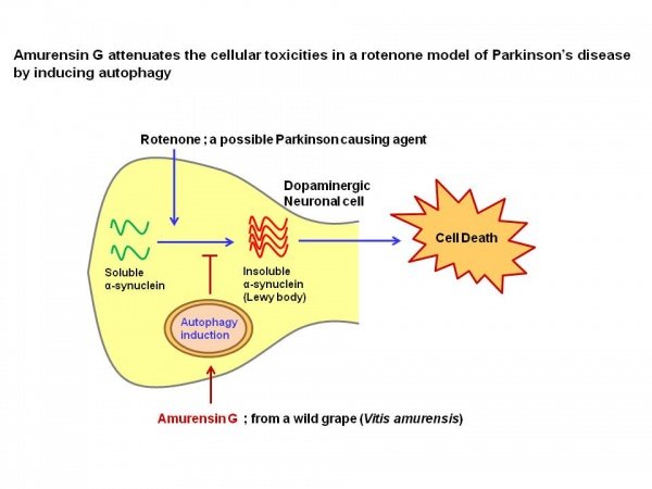
- Paraquats mechanism of action is the production of reactive oxygen species, intracellular molecules that cause oxidative stress and damage cells.
- Rotenones mechanism of action is disruption of the mitochondria, the component of the cell that creates energy for cell survival.
Interestingly, both mitochondrial dysfunction and oxidative stress are common themes in our general understanding of what causes death of nerve cells in PD.
Read Also: Does Sam Waterston Have Parkinsons
Rural Living And An Increased Risk Of Pd
In the 1980s, studies were conducted that showed that early-age exposure to a rural environment as well as exposure to well water were associated with development of PD later in life. Subsequently, multiple additional studies looked at these questions. The studies are mixed in their conclusions, but overall the evidence supports associations between increased PD risk with each of the following:
- farming as an occupation,
- well water drinking, and
- living in a rural area.
Of course, all these categories are inter-related, since farmers live on farms in rural areas, are exposed to farm animals, are more likely than urban dwellers to drink well water and use pesticides. The studies were attempting to tease out why rural environments increased the risk of PD. Do only those who actually farm have an increased risk or is it enough to live on a farm? Is pesticide exposure the reason for the increased risk? Well water exposure? Exposure to farm animals? Or is it another element of rural life?
In the end, epidemiologic data supports the assertion that each of these elements increases the risk of PD. Of note, all of the increased risks in these studies are small on the order of 1.5-2 times the risk of the general population.
Recommended Reading: On Off Phenomenon
Western Blot Analysis Of Nrf2 Ho
Total protein from the frozen striate was extracted by the aid of The ReadyPrepTM protein extraction kit . Assessment of protein content was carried using the Bradford protein assay kit from Bio Basic Inc. . Protein from each brain sample, 20 g, was denaturized through boiling at 95°C for 5 min in buffer . Then, protein was loaded in polyacrylamide gels that were relocated to a polyvinylidene difluoride membrane. After that, the PVDF membrane was blocked by tris-buffered saline with Tween 20 and 3% bovine serum albumin for 60 min at room temperature. PVDF membranes and primary mouse-specific antibodies of HO-1 , Nrf2 , AMPK alpha-1 , FOXO3 , thioredoxin , cleaved caspase 3 , and vascular endothelial growth factor , and -actin were incubated overnight at 4°C and rinsed with TBST. Then, incubation was performed in horseradish peroxidase-conjugated secondary antibody at room temperature for 60 min. The blot was rinsed with TBST80 and detected via chemiluminescent kit . Film bands were captured by a CCD camera-based imager . Band intensity of the target proteins were quantified after normalization by -actin using Image-J1.52p .
Determination Of Cell Viability By Mtt Assay

SH-SY5Y cells were seeded in 96-well plates and incubated overnight. Rotenone were freshly dissolved in DMSO and added to the cells at a final concentration of 0.5% DMSO for each experiment. Danshensu dissolved in DMEM was added at 2h prior to the rotenone exposure. To determine the safety of Danshensu and the toxicity of rotenone, cells were treated with different concentrations of Danshensu or rotenone for 24h. To evaluate the neuroprotective effect of Danshensu, SH-SY5Y cells were pretreated with Danshensu for 2h and then challenged with rotenone for 24h. The cells were incubated with 5mg/mL MTT for 4h at 37°C in the dark. The medium was removed carefully after the incubation of MTT. The formazan crystals were dissolved in 200L DMSO and the absorbance of formazan reduction product was measured by microplate reader at 595nm. Cell viability was expressed as values relative to the control with a control value of 100%.
Don’t Miss: Prayers For Parkinson’s Disease
Immunohistochemistry For Tyrosine Hydroxylase
Tyrosine hydroxylase immunostaining revealed strong cytoplasmic staining in the vehicle control group in the SNpc region . However, the rotenone control group showed reduced and irregular staining . Images from the mice groups treated with MTF showed greater and regular area for staining . Analyzing the number of TH-stained neurons showed the greatest number in the vehicle group, and this number was reduced significantly in the rotenone control group. Staining areas increased dose-dependently in mice receiving MTF . Similar results were obtained from striatal immunostaining TH staining was strong and regular in the vehicle group but decreased and was sporadic in the rotenone control group . Mice groups treated with MTF showed gradual enhancement in staining .
Analysis of the striatal stained area indicated a significant decrease in the rotenone group versus the vehicle group , and dose-dependent increments were observed in mice groups treated with MTF . On the other hand, the mice in groups 5 and 6 treated with the vehicle + MTF 100 or 200 mg/kg showed intact SNpc neurons similar to those observed in the vehicle control group. Importantly, the number of the TH-positive neurons in groups 5 and 6 was not significantly different from that observed in the vehicle group .